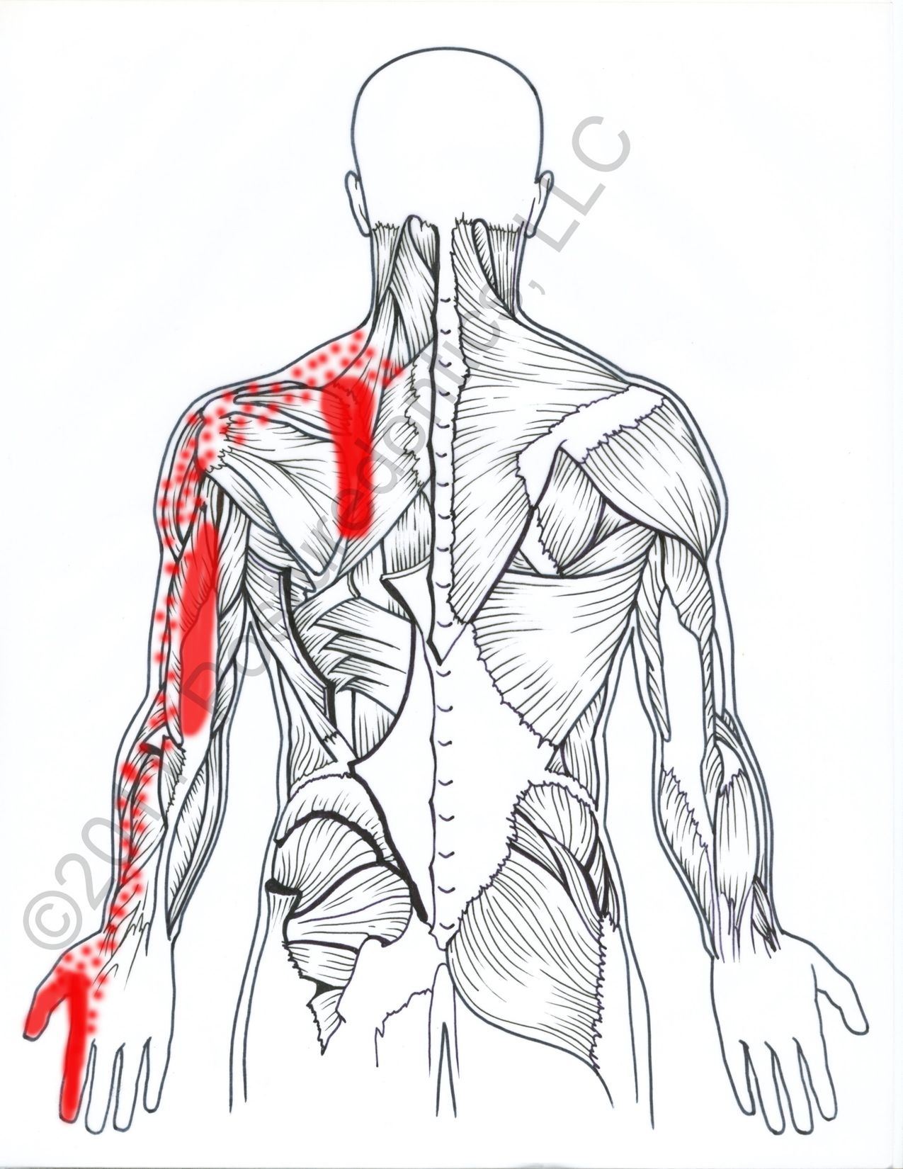
Left Upper Back Anatomy : Chest Wall Lumps Rib Injury Clinic : The upper back is often referred to as the thoracic spine and is generally.. Longissimus and quadratus lumborum the longissimus (red, in the image above) are located between spinalis and the iliocostalis muscles. The axial muscles are grouped based on location, function, or both. This system reflects the bones of the skeleton system, which are also arranged in this manner. It is like that for several reasons, all of which you can understand by looking at the anatomy of the thoracic spine. License image the deltoid, teres major, teres minor, infraspinatus, supraspinatus (not shown) and subscapularis muscles (not shown) all extend from the scapula to the humerus and act on the shoulder joint.
This plexus splits into nerves that carry sensory messages and. It runs from the neck to the upper back. Anatomy of human body organs 12 photos of the anatomy of human body organs anatomy human body organ systems foldable, anatomy of human body and its functions, anatomy of human body drawing pdf, anatomy of human body parts, anatomy of human body quiz, human anatomy, anatomy human body organ systems foldable, anatomy. After, behind, following, toward the rear distal: These two terms, used in anatomy and embryology, describe something at the back (dorsal) or front/belly (ventral) of an organism.
Upper Back Pain Anatomy Of The Back The Pain Center Pain Management Care from www.2-boots.com The dorsal (from latin dorsum 'back') surface of an organism refers to the back, or upper side, of an organism. These sections are cervical (neck), thoracic (upper and middle back), lumbar (lower back), and sacrum (tailbone). Near, closer to the origin dorsal: Pain in the upper left abdomen causes and conditions. The left upper quadrant (luq) includes the lower left ribs, stomach, spleen, and upper left area of the transverse colon. It runs from the neck to the upper back. Away from, farther from the origin proximal: In the upper back region, the trapezius, rhomboid major, and levator scapulae muscles anchor the scapula and clavicle to the spines of several vertebrae and the occipital bone of the skull.
The human spine is composed of 4 sections of vertebrae.
It runs from the neck to the upper back. Upper back pain is typically due to one of the following: Sometimes, pain felt on the left side of the middle back can be coming from a nearby organ. The upper back is often referred to as the thoracic spine and is generally. Related posts of anatomy of the back organs anatomy of human body organs. The cervical spine protects the nerves connecting to the brain, allowing the head to move freely while supporting its weight. Toward the bottom, toward the belly superior: Spinal nerves l1 through l4 converge to form the lumbar plexus. There are three sets of iliocostalis muscles: Where is my left upper quadrant? Longissimus and quadratus lumborum the longissimus (red, in the image above) are located between spinalis and the iliocostalis muscles. The axial muscles are grouped based on location, function, or both. They originate from the vertebrae and insert into the scapulae.
Because the radial nerve wraps around the humerus bone, it can be stretched or torn when the humerus bone is broken. The left upper quadrant is the area of the abdomen that is on the left side region of the navel and extends up to the left rib cage. Rest, ice, compression and elevation. In other cases, the pain may be unrelated to your back. The rhomboid muscle is activated as you bring and squeeze your scapula or shoulder blades back and together.
A Mysterious Upper Back Pain Syndrome In Dental Professionals from cdn.sanity.io Formed by the merging of spinal nerves c5 through t1, this plexus branches into nerves that carry sensory messages and provide motor control to the muscles of the arm and upper back. 1) in the cervical area (iliocostalis cervicis), 2) in the upper back or thoracic area (iliocostalis thoracis), and 3) in the lumbar area (iliocostalis lumborum). Causes of pain in the upper left abdomen can be varied. Look down at your tummy, and mentally divide the area from the bottom of your ribs down to your pubes into four quarters. The upper left back is the region to the left, below the neck (cervical spine), and above the lower back (lumbar spine). Toward the bottom, toward the belly superior: License image the deltoid, teres major, teres minor, infraspinatus, supraspinatus (not shown) and subscapularis muscles (not shown) all extend from the scapula to the humerus and act on the shoulder joint. It consists of seven vertebrae.
Longissimus and quadratus lumborum the longissimus (red, in the image above) are located between spinalis and the iliocostalis muscles.
This point of termination is called the conus medullaris, 7 from where the spinal nerves descend down. Take the upper extremity anatomy quiz and learn more about the bones, joints, muscles and vessels of the upper extremity! Knowing the anatomy of the thoracic back can help to locate the source of back rib pain on your left side. These two terms, used in anatomy and embryology, describe something at the back (dorsal) or front/belly (ventral) of an organism. The nerve gives function to the triceps muscles on the back of the arm to straighten the elbow. Muscles in your upper left back and shoulder that you could potentially strain include your rhomboid, trapezius, deltoid or infraspinatus. This system reflects the bones of the skeleton system, which are also arranged in this manner. Sometimes, pain felt on the left side of the middle back can be coming from a nearby organ. Because the radial nerve wraps around the humerus bone, it can be stretched or torn when the humerus bone is broken. Spinal nerves l1 through l4 converge to form the lumbar plexus. Pain in the upper left abdomen causes and conditions. Both the deltoid and the trapezius are firmly attached to … Anatomy of human body organs 12 photos of the anatomy of human body organs anatomy human body organ systems foldable, anatomy of human body and its functions, anatomy of human body drawing pdf, anatomy of human body parts, anatomy of human body quiz, human anatomy, anatomy human body organ systems foldable, anatomy.
Take the upper extremity anatomy quiz and learn more about the bones, joints, muscles and vessels of the upper extremity! The skeletal muscles are divided into axial (muscles of the trunk and head) and appendicular (muscles of the arms and legs) categories. These sections are cervical (neck), thoracic (upper and middle back), lumbar (lower back), and sacrum (tailbone). There are three sets of iliocostalis muscles: The cervical spine supports the weight and movement of your head and protects the nerves exiting your brain.
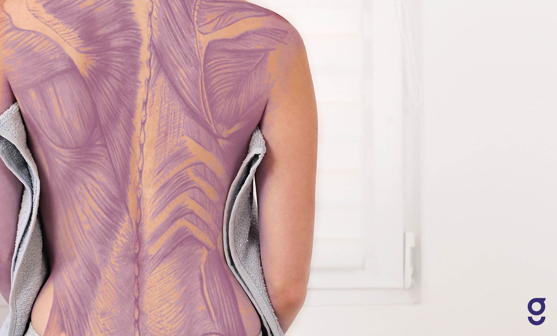
Back Muscles Anatomy Of Upper Middle Lower Back Pain In Diagrams Goodpath from images.ctfassets.net You should consult your physician for pain that doesn't resolve and to receive a definitive diagnosis. In other cases, the pain may be unrelated to your back. In front of, front posterior: Both the deltoid and the trapezius are firmly attached to … The dorsal (from latin dorsum 'back') surface of an organism refers to the back, or upper side, of an organism. Toward the bottom, toward the belly superior: Upper back pain is typically due to one of the following: This plexus splits into nerves that carry sensory messages and.
Knowing the anatomy of the thoracic back can help to locate the source of back rib pain on your left side.
These descending spinal nerves resemble a horse's tail and are called the cauda equina. The cause may be poor posture (such as forward head posture) or any type of irritation of the large back and shoulder muscles, including muscle strain or spasms. In the upper arm the radial nerve wraps around the back side of the humerus bone. The axial muscles are grouped based on location, function, or both. Rest, ice, compression and elevation. Longissimus and quadratus lumborum the longissimus (red, in the image above) are located between spinalis and the iliocostalis muscles. Organs like the kidneys or pancreas can cause pain that spreads to. The left upper quadrant (luq) is a section of your tummy (abdomen). Related posts of anatomy of the back organs anatomy of human body organs. The lumbar and sacrum region make up the bone of the lower back anatomy. The dorsal (from latin dorsum 'back') surface of an organism refers to the back, or upper side, of an organism. In the upper back region, the trapezius, rhomboid major, and levator scapulae muscles anchor the scapula and clavicle to the spines of several vertebrae and the occipital bone of the skull. The organs found in the left upper quadrant are the left sections of the liver and kidney, adrenal gland, spleen, stomach, pancreas, colon's splenic flexure and lower portion of the colon.
This system reflects the bones of the skeleton system, which are also arranged in this manner upper back anatomy. This plexus splits into nerves that carry sensory messages and.


0 Komentar